P3D Biology 1
Provides a fantastic journey through the human body and its systems. The 3D models in virtual reality are divided into three parts: muscles, skeleton and internal organs. In those cases, in which microscopy is important, the models show both the macroscopic and also the microscopic part: all in 3D. For example, the human eye and the vision are shown in detail: from the structures of the eye to the retina tissue with their rods and cones. And we can follow the human fetal development from its 18th day to its 180th day of life. The module is completed with 3D animation videos showing how the human body systems work.
Models
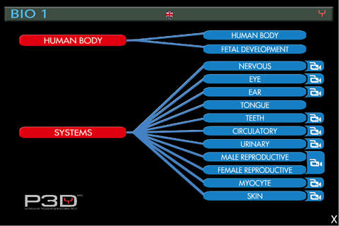
BIOLOGY 1
HUMAN BODY
• Human Body
• Fetal Development
SYSTEMS
• Nervous
• Eye
• Ear
• Tongue
• Teeth
• Cardiovascular
• Urinary
• Male Genital
•Female Genital
• Myocyte
• Skin
ANIMATIONS
• Nervous System: Shows the conduction of nervous impulses, synapses and the voluntary movement of taking a glass to the mouth.
• Eye: shows the accommodation of the crystalline lens and the formation of the image on the retina, standing out cones and rods.
• Ear: shows the action of the tympanum and that of the ossicles of the middle ear, and, additionally, it shows how these organs act for the equilibrium and balance of the body.
• Teeth: Shows the complete adult human teething. It is possible to view the inner side of the teeth and the cariogenic action.
• Cardiovascular: shows the heart during systole e diastole, the long and the short circulation with the pathway of arterial and venous blood.
• Urinary: Shows the arrival of blood through the renal artery and its passage through the nephrons, with consequent filtration and formation of urine.
• Male and Female Genital: shows the spermatogenesis and sperms leaving the male genital organ. Shows the ripening of the follicle, ovulation, fertilization and initial stages of the embryo development
• Myocyte: Shows a muscle fiber with the actin and myosin arrangement during muscle contraction.
• Skin: Shows the epidermis and the dermis with all of their sensorial structures, pilous follicle, sebaceous and sudoriparous glands.


